Showing posts with label cases. Show all posts

Saturday, February 3, 2018

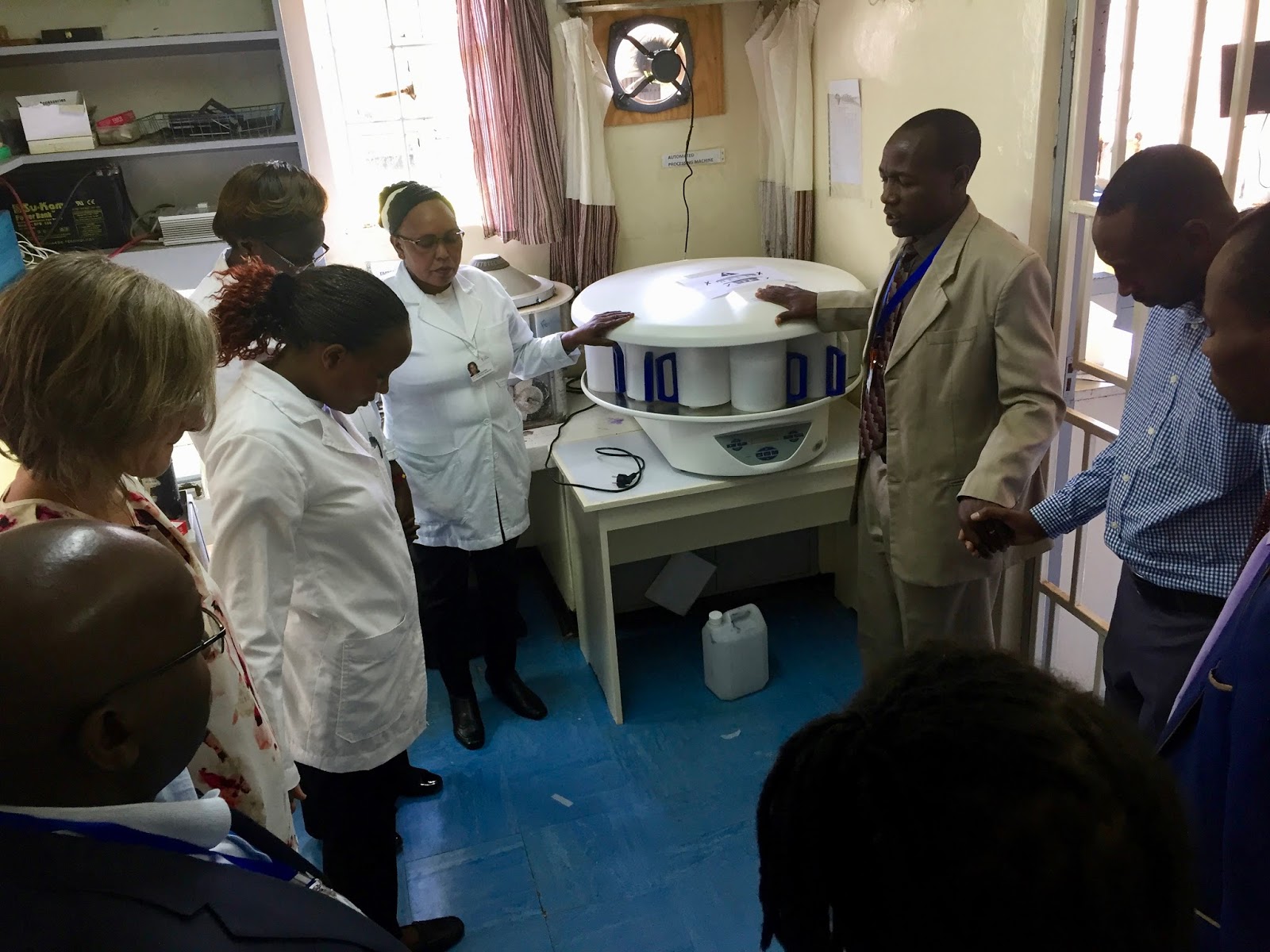
The new processor!

During my rotation, the most exciting change in the laboratory was getting a new tissue processor.  For those who don't know much about anatomic pathology, making an accurate diagnosis for patients relies extensively on a technique that hasn't changed much since the 19th century:  hematoxylin and eosin stained histologic sections of human tissue specimens.  These are cut from fixed and processed tissue embedded in paraffin wax by histotechnologists.  The processing part requires a lengthy procedure, often done overnight.  It includes fixation of fresh tissue in formaldehyde solution (formalin), dehydration of the tissue with alcohols, clearing with xylene, and embedding the tissue in paraffin wax in a "block."  While the processing used to be done by hand long ago, there are many steps using solutions and solvents required in a modern tissue processing program, and clinical workflows tend to have a large number of specimens, so an automated machine is the norm.  Here is a decent source (from a big company...) that explains it for those who may not have much exposure: https://www.leicabiosystems.com/pathologyleaders/an-introduction-to-specimen-processing/

The old processor is on the right
Getting the the new one in the door.
Plug it in!  This model - the STP 120, can cost ~$25,000 if purchased new at full price per my online research, and it seems to be a cheaper alternative.
Dedication and prayer for the processor with hospital officials!  This usually does not happen at UWMC.
Lifting up the hood and showing the rotating baskets and solution containers or retorts.
Something you might have noticed with this processor (and the old one), if you've been around them before, is it looks a bit different.  Here's more what you might expect (from the Leicabiosystems site):Picture of a modern enclosed tissue processorYou are probably used to seeing an enclosed tissue processor such as this one.  All of the tissue is in one container (or retort) and the solutions are heated as needed and pumped into and out of the retort.  In the "dip and dunk" processors that we use in the Kijabe lab, the specimens are moved from solution to solution in a rotating carousel fashion.  These models don't have as many features for increased temperatures and pressures to reduce processing times and increase efficiency as the enclosed processors do, but they are less expensive and easier to maintain, making them a good choice for Kijabe's pathology lab where turnaround time is less valued and cost, reliability, and maintenance issues are more important.  I had never seen the dip and dunk design before, and I'm glad I got to help install and dedicate this shiny new piece of equipment!

Hooray for the new processor!  Best wishes, and may it serve the Kijabe lab well for many years.

Wednesday, January 31, 2018

And the winner is:

... a Kijabe alum, Josh Lieberman.  Thanks for submitting the answers, Josh!

In addition to the Strandjord-Clayson award he got today (CONGRATULATIONS!!!!) for his laboratory medicine achievements, he also will be awarded the coveted Tab Stealing Monkey Award for skill in Kijabe related histopahtologic diagnostic skills.


I'm putting the answers below the original post in small font with explanations.


Wednesday, January 24, 2018

Quick trio of cases! Can you diagnose them all?

Anatomic pathologists and pathology enthusiasts - check out these cases.  These are fairly routine cases from Kijabe.  Can you get them?  First to email me (Thomas Long) with all 3 correct diagnoses will be recognized on the blog.  Differential diagnoses will not be accepted.  One guess per case.

Case 1:  The patient is a 28 year old Kenyan man with HIV.  He complained of dysphagia and the oesophago-gastro-duodenal (OGD) endoscopy showed a mass.  Biopsy is pictured below.  What is your diagnosis?


Case 2:  A 36 year old Kenyan woman presents with a cervical mass.  Radical hysterectomy with lymph node dissection is performed.  Mass and lymph node are pictured.  Diagnosis?


Case 3:  Now for some cytology.  A 45 year old Kenyan man presents with a left inguinal mass.  FNA is performed.  Diagnosis?


ANSWERS:

1.  This nodule in the esophagus of someone who has HIV and is likely immunocompromised contains numerous foamy macrophages and especially on highest power (oil immersion, 100x) clearly shows intracellular organisms morphologically suspicious for histoplasmosis.

2.  The pictures are intentionally from lower power.  There is a solid-appearing invasive carcinoma.  The most common diagnosis and the one that comes to mind first would generally be squamous cell carcinoma, but the lesson here is that not all that is solid is squamous.  If you look carefully, there are gland-forming lumens, most easily identified on the lymph node metastasis.  Additionally, the cells are rounder and less angulated than typical squamous cells, and there is no keratinization or other obvious squamous feature other than somewhat eosinophilic cytoplasm.  Here is a higher power view of more obvious gland formation:


3.  This cytologic specimen is first positive for a epithelioid to spindled, cohesive malignancy.  But the nuclear features (round with some prominent nucleoli) and especially the abundant cytoplasmic pigment with a somewhat granular quality, are highly suspicious and essentially diagnostic of melanoma, in this case metastatic to an inguinal lymph node.  This patient had a primary on the foot, and this likely falls into the category of acral melanoma, which is the most common form of melanoma in African Americans, and probably also Africans.  We saw a couple of cases (both fairly advanced), and that's about all the skin cancer we saw during my rotation.

Monday, January 23, 2017

So what is cancer?


So what is cancer, anyway?
By Josh & Rochelle

As Pathologists we deal with this question every day and while this might seem like a straightforward question, it requires some careful consideration.  Like any good question, trying to answer “what is cancer” leads us to a series of additional, equally interesting questions.  Before reading on, take a minute and fix a definition of cancer in your mind.  

Got one?  Ok, good. 

Cancers – and I emphasize the plural, because there are a great many different diseases that are all “cancer” – are a type of something that we generically call neoplasms (neoplasia simply means “new growth”). But not all neoplasms are cancers – so what’s the difference? 

I think most people would agree with us in saying that neoplasm is an uncontrolled, clonal proliferation of cells carrying a mutation in their genetic material (a “somatic mutation”) and that this uncontrolled growth may damage the patient.  That’s a good start, but let’s dig a little deeper.  This process starts from some precursor cell with the potential to divide that acquires genetic changes.  These changes (mutations) allow the cell to divide and give rise to daughter cells that keep dividing, all ignoring the usual signals that tell them to stop and/or die when they should.  We usually talk about “two hits” being required for this process called “transformation.” 

So when does a neoplasm become cancer?  We argue that “cancer” identifies a neoplasm that is locally destructive and/or can metastasize (spread to a different site).  It is these two features – local destruction and metastatic disease – that are clinically so very dangerous for patients.

Pre-Cancer vs Cancer
The body has stem cells to replenish damaged or shed normal cells at the end of their lives.  The gut and skin, for example, routinely and rapidly turn over new cells in predictable, genetically-regulated cycles that ensure we have an intact covering over our body to prevent infections, retain moisture, and absorb nutrients.  But the body has stem cells ready to replenish essentially every part of itself! 
Overtime, these stem cells can acquire transforming mutations either by chance, because the person has inherited a defective copy of the genetic proof-reading machinery, or due to an infection.  These cells can start to grow, ignore boundaries and stop signals, and produce a change that we can see with our eyes or a microscope.  These growths are also neoplasms!  Often, the body’s immune system can find a way to kill these misbehaving cells – especially when helped by a doctor cutting out this neoplasm. 

When neoplasms occur in the colon, the gastroenterologist typically sees polyps.  In the cervix (think HPV infection), a gynecologist finds discolored or eroded tissue.  In the breast, we might call this “ductal (or lobular) carcinoma in situ.” In the colon we call these “adenomas” and in the cervix we call this “intraepithelial neoplasia.”  The difference between each of these three processes is only in the spelling. These processes are all neoplasms arising from the lining of the body (mucosal surface or “epithelium”) and at the time of detection have not “invaded” into the underlying tissue.  Invasion is a complex process that requires eating and/or squeezing through some sophisticated networks of proteins that the body builds to separate its different compartments. 

The above examples – adenoma, carcinoma in situ, and intraepithelial neoplasia – are all types of “pre-cancers.”  This means they all have the potential to invade, but right now they are contained.  It’s like a misbehaving child who is grounded and stays in the house.  Catching these lesions early is precisely the goal of screening programs, like getting a Pap smear.  If left to their own devices, these lesions may (many do not) invade and from their spread throughout the body and wreak havoc. 
Examples in Pictures:
Colon: Normal, Intraepithelial, and Invasive 
Panel A shows normal colonic epithelium.  Part B shows an overgrowth of cells confined to the surface, an intra-epithelial neoplasm.  Part C shows glands running around beneath the surface; this is an invasive neoplasm (cancer), or in this case, an adenocarcinoma which indicates the cancer arose from gland-forming epithelium.  Panel A & C are from a case in Kijabe.
Breast: Intraepithelial and Invasive
Panel A shows a hypercellular duct, but all this new cells growth is contained within the duct.  If you look closely, you can see thin, spindled cells lining the duct and separating the glandular cells (in the duct) from the pale, pink, whispy supportive tissue (stroma).  Panel B is quite different, with duct-like structures invading throughout the stroma.  Unfortunately I don't have a great picture of normal breast epithelium to share, but it's basically a much smaller, less cellular version of Figure A with some glands hanging off the end.  Both of these figures are from a case we had recently in Kijabe.
Cervix: Normal, Intraepithelial, and Invasive
Panel A shows essentially normal epithelium (the layered, pink part that looks kind of like a basket weave at the "top" of the tissue in the image).  Panel B shows bigger, irregular cells in the epithelium with loss of the basket weave appearance.  Panel C shows destruction of the epithelium with those bigger, uglier cells invading the pink stroma in clusters and single cells. All of these images are from cases here in Kijabe.
Benign vs Malignant
These two terms, “benign and malignant,” are highly fraught and used quite imprecisely.  Benign means innocuous, but is an adenoma in the colon really innocuous?  How about ductal carcinoma in situ (DCIS) of the breast?  What about cervical intraepithelial neoplasia (CIN)?  None of them have invaded, all have the potential to invade and to go on and kill the person, but not all will progress. 
Many people – clinicians included – might call a colon adenoma “benign”, while deciding that DCIS is malignant – but biologically they are the exact same process.  Even the major US initiative that collects data on neoplastic diseases, the Surveillance, Epidemiology, and End Results program (SEER) run by the National Cancer Institute, includes DCIS as cancer but does not count colonic adenomas (non-invasive).  Many of us in pathology and the basic sciences object to this; why this distinction is made is an open question. 

Let’s ask the question one more time, but in a different way.  Are invasive colon cancer, invasive breast cancer, or invasive cervical cancer benign or malignant?  Of course they are malignant!  So perhaps the best thing we can say is that the pre-cancerous lesions “have the potential to invade and become cancers.”    

I'll add that "benign" doesn't necessarily mean "safe no matter what."  A couple of non-epithelial neoplasms demonstrate this nicely.  A meningioma is a "benign" brain neoplasm that doesn't infiltrate like a glioma and doesn't metastasize, but if in the wrong location can be fatal.  Similarly, uterine fibroids are a smooth muscle neoplasm called a leiomyoma. Usually the worst that accompanies a fibroid is pain or heavy periods, but someone with heavy bleeding from a fibroid could die from blood loss or secondary infection.   

Conclusions
I hope this has been a useful discussion of the terms “cancer,” “neoplasm,” “benign,” and “malignant.” These terms and diagnoses like those above carry enormous emotional impact for patients.  What we call these entities doesn’t necessarily reflect their biological behavior. For example, many people would consider DCIS to be “cancer” but wouldn’t be particularly perturbed by the diagnosis of a tubular adenoma in the colon.  However, they are fundamentally the same biological process. 

Wednesday, January 18, 2017

What is among us?

File under look-a-likes.
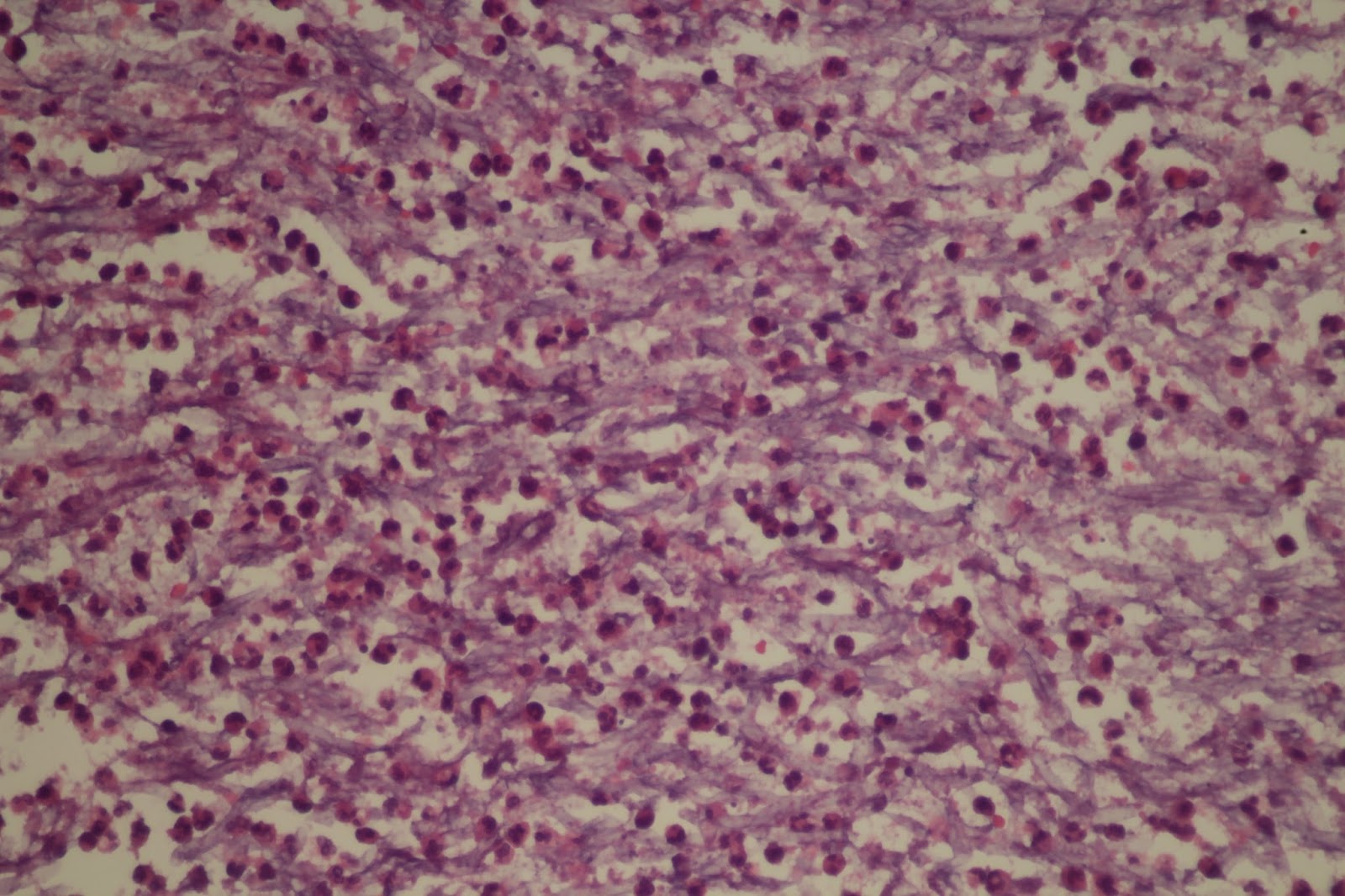
Histoplasma?  Platelets?

We went with platelet clumps.

Friday, January 13, 2017

Gross Dx Challenge - Answers & Micro Images!

We previously posted a diagnostic challenge:
12 year old female with an abdominal/liver mass consisting of lots of little cysts.
Did you figure it out?

If you weren't sure, here are some micro images should help and that prove our diagnosis!

That's right, it's a hydatid cyst. These are infections by the tissue-infectious Cestode or tape worm Echinococcus. [The CDC has a fantastic set of websites on parasitic infections, including lots of detailed information on organisms and life cycles]. Echinococcus is usually transmitted by dogs to humans via fecal-oral contamination. The worms tend to set up shop in the liver, creating a cystic mass that gets filled with daughter cysts containing parasite larvae (seen in the gross photos, linked above). Patients present with vague abdominal pain and the treatment is surgical resection of the mass.
For more information about some of the many, many parasites in the world, I strongly recommend reading the book Parasite Rex.

Brief follow-up: my colleague and friend from home correctly asked, "Hey, if this thing ruptured in the patient, did she get really sick?"  He, being a skill physician, asked this in a more medical manner and wisely so because hydatid cysts CAN rupture and result in a severe allergic reaction (anaphylaxis) secondary to a big parasite burden.  But only sometimes.  This little girl DID have an intrabdominal rupture of the cyst (and the surgeons and I agree it did not smell nice), but was fine!  She is now being discharged and is a happy little camper, although she is on 6 months of anti-parasitic drugs.  I'll try to post an intra-operative picture soon.

Tuesday, January 10, 2017

Gross Diagnosis Challenge!

This lesion should be a pretty straightforward diagnosis.  I got the contents of a ruptured cyst, complete with fragments of (some non-viable) cyst wall.  I don't have micro images, yet.  In the meantime, can you make a gross diagnosis?

Hint: This should be interesting for Lab Med folks, too!
There were LOTS of these little "marbles" plus the remnants of the cyst wall.
Close up of one of the "marbles" with cloudy flocculant stuff in it.

Here's an artsy B&W image.


All photo credits: Nicole Lieberman, PhD and all-around badass.


Sunday, January 8, 2017

Kijabe Hospital & Access to Health Care

Kijabe Hospital  has 365 beds, including an ICU, operating theater, men's & women's wards, pediatric, ob-gyn, ENT, Infectious Diseases/HIV, and outpatient services.  The hospital is part of a larger network of outlying clinics, draws from a large geographic catchment zone, and offers pathology referral services - a critical and severely understaffed area of medicine in Kenya. We see about 3500 pathology cases every year. The last few weeks have been complicated by a national strike among doctors and nurses in public hospitals.
NPR - Doctor's Strike in Kenyan Public Hospitals

Okay: the hospital here provides important services to a large number of people.  But how hard is it to get here? 

When I walk into work every day, I pass through patient waiting areas.  This morning I was pulled aside by a young man, about my age (early-to-mid 30s), who was here with his wife and young son.  The man was able to explain that his son had "water in his head" (hydrocephalus).  The patient is a 2 month old, former 34 week permature infant with a birth complicated by NICU stay and hydrocephalus. This is apparently a common problem here due to folate & B12 deficiency.

The boy had been evaluated in the emergency room overnight, but the family was still waiting. The father asked for help getting seen by the doctors as he was understandably worried about his son and concerned that "nothing had been done for [the boy]." We talked for a few minutes. I established that the boy had already been evaluated, which meant there wasn't an acute emergency. The father politely responded that, to him as the boy's father, this was an emergency. I did my best to reassure him that the doctors would be out after their morning rounds, probably about 0900, and that I would check back around 0930 or 1000 to see if he had been seen at that point. I shared that I was new here, and mostly worked in the labs, but would do what little I could.

As I talked with the man in the waiting area, I was struck by his and his family's patience. They had traveled 9hrs to get to the hospital and slept in the ED. They have no car and transportation can be expensive.

Travel Time: Kenya vs US and other high-income countries
American hospitals also have long ED wait times and, for many rural communities, access to specialized or tertiary care centers is a major challenge. Nine hours struck me as an almost inconceivable amount of time for the majority of areas in the developed world. At home, we have systems of ambulances, helicopters, and learjets to transport critically ill patients. So how does this family's experience compare to the average travel time to tertiary care centers in the US & other high-income countries?

In North England, median travel time was ~28min with outliers out to 90-120min.  Most travel was by private car.  Ref 1

In the US, ~80% of patients live within 1h of access to an institution that can provide cardiac catheterization (PCI) in the event of a heart attack. Ref 2, 2006 data

As of 2001 data, ~86% of Americans traveled by car (or private vehicle) for their >5 BILLION yearly medical & dental trips. For all visits, rural trips averaged 27 min (17.5 miles) compared to 20.7 min (8.3 miles) for urban residents.  ~8% of US patients traveled more than 30miles and 28.5% of trips took >= 30min.  Not surprisingly, the burden fell overwhelmingly on rural residents. Although other patterns of inequity in access to care were also observed - non-white patients were >5x more likely to rely on public transit/walking; African Americans had on average 50% greater travel times than whites (29min vs 20min) - but these are discussions for a different post.

Further reading on rural care, methods, and access in Idaho (which has some of the lowest numbers of practicing physicians) see this article.

Case of Week 1

UPDATE!
With input from our Peds Path collaborators, we have diagnosed this lesion as a mesenchymal hamartoma  of the liver (MH). An important consideration for this lesion is that an undifferentiated embryonal sarcoma (UES) can arise within this hamartomatous process. This is indeed what we identified in this case. As you'll see below (40x objective, HPF), we have found areas with significant cytologic atypia and multiple mitoses (we maxed out at 5 per HPF).
Multiple mitotic figures are present in this area of atypical stromal cells.
Significant stromal cell atypia, getting towards anapastic.
Diagnosing the UES arising within the MH is important as survival has traditionally been poor (~37%), although recent reports suggest 70-100% survival may be possible.  Unlike mesenchymal hamartoma, surgical excision alone is not curative.  Ref 1

As usual, pathologic diagnosis is really critical for this lesion as MH and UES (or UES/MH) are quite difficult to distinguish pre-operatively. Ref 2

Sadly, although we have provided the diagnosis and our patient can get into definitive care, given the resources in rural East Africa I suspect their odds of survival are considerably lower than 100%.


Case of the Week: Week 1
 We get a broad mix of pathology specimens here at Kijabe Hospital, including some rare entities and a some cases common here but unusual in the US.  Nicole will be posting more extensive information on the sorts of cases and how these patterns have changed over time (she's currently deep in a large data set).

Today I want to share the most challenging case we had in our first week.  We have not yet put a final diagnosis on this and comments are welcome.  We have requested a digital pathology consultation from our Pediatric Pathology colleagues back home.

The specimen is a liver resection from a 7 year old.  We were provided with limited clinical history but we were told that the liver ruptured in the patient.

Gross Examination
Gross image.  Note the site of rupture (top right).  Photo Credit: Nicole Lieberman.

Gross image.  Cross section.  Multiple pale, firm areas, as well as areas of hemorrhage and probable necrosis.  At right, glassy-green mucoid areas also visible.  Photo Credit: Nicole Lieberman.


The 2300 g liver is irregularly enlarged, nodular, with pale and firm areas as well as foci of central necrosis. Several mucoid foci are present. There is essentially no preserved normal liver parenchyma. The gallbladder is grossly unremarkable with smooth, velvety, green-brown mucosal surface and no ulcers or firm areas. An unremarkable 1 cm hilar node is also unremarkable.

Microscopic Examination
Low power (4x objective) view of nodular areas.  Mixed densely cellular areas as well as possibly myxoid foci.  Possibly entrapped ductules and vascular structures.  (The dark purple is pigment precipitate).

Low power (4x objective): At right there appear to be more normal trabecules of hepatocytes.  At left there are pale areas with distinctly different cellular growth patterns. 

Low power (4x objective) view of mucoid areas seen on cross section. Multiple dilated bile ductules in a totally abnormal arrangement with luminal necrtoci debris. No epithelial atypia is present. Dilated blood vessels are also visible. 
High power (40x objective) view of paler areas.  Cells here appear to have large, possibly vacuolated cytoplasm with moderately enlarged nuclei.  The nuclei appear to have inclusions/vacuolations. 

40x: Entrapped bile ductules adjacent to hepatocytes (right) with scattered abnormal cells, similar to those seen in above photomicrograph.

 40x: Densely packed atypical cells with entrapped vascular structures.
Final Diagnosis: Pending.


Suggested Reading:
Archives of Pathology - HMH
Pediatric Liver Masses - Path & Rads Correlation
Hepatoblastoma - Path Outlines Review
Fibrolamellar HCC - Path Outlines

Wednesday, January 20, 2016

Infectious Disease Edition

20 January 2015

Here are a few cases from the last week with a particular theme...

Case #1
History: 
29-year-old male with history of multiple courses of treatment for Mycobacterium tuberculosis. He now presents with moderately severe hemoptysis. 
Gross: Partial resection of upper lung lobe. There is a cavity lined by apparent bronchial mucosa that is filled with an aggregate of friable brown material, measuring 3 x 2 x 2 cm. 


Lung resection specimen with cavitary lesion filled with brown, friable material


Low power image of lung and friable mass
High power view of lung and friable mass
Medium power view of mass


High power view of mass

Case #2
History: 19-year-old female with abdominal pain and distention, dilated colon, congested liver with bile-stained fluid. Clinical concern for hepatitis.
Gross: 2 cm liver biopsy.

Low power view of liver
Ziehl-Neelsen stain

Ziehl-Neelsen stain

Ziehl-Neelsen stain  
Case #3 (Repeat from previous post with better images)
History: 26 year-old female with HIV (last CD4 count 250 cell/uL) and chronic sacral/gluteal wound that began as a small vesicle.
Gross: Punch biopsy of skin, at the interface of the wound and normal-appearing skin.

Photograph of sacral/gluteal wound


Low power view of normal skin and lesion

Medium power view of lesion
High power view of lesion

Consider the diagnoses and I'll update the post with our interpretations after we return from an attempt on Point Lenana, Mount Kenya (16,355 ft)!

And hopefully the weather looks more like this....


Road to Kijabe on a training hike

And less like this....


Monsoon hail storm on the crater rim of Mount Longonot (9,108′). Rochelle is in the blue and white coat. Phillip is well-protected behind the guy in the red shirt (white pant leg).


Our interpretations:
Case #1: Left lung, partial lobectomy: Fungal ball consistent with that caused by Aspergillus sp. (aspergilloma, 3 cm) surrounded by granulation tissue, necrotic tissue, and respiratory mucosa; no definitive invasive fungus identified.
Case #2: Liver, biopsy: Granulomatous hepatitis with rare acid-fast bacilli identified on Ziehl-Neelsen stain, consistent with extra-pulmonary Mycobacterium tuberculosis infection. 
Case #3: Skin, sacral and gluteal, biopsy: Ulcerated skin with neutrophilic inflammation and epithelial cells with probable viral inclusions, consistent with herpes simplex virus infection.