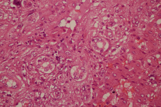
With input from our Peds Path collaborators, we have diagnosed this lesion as a mesenchymal hamartoma of the liver (MH). An important consideration for this lesion is that an undifferentiated embryonal sarcoma (UES) can arise within this hamartomatous process. This is indeed what we identified in this case. As you'll see below (40x objective, HPF), we have found areas with significant cytologic atypia and multiple mitoses (we maxed out at 5 per HPF).
| Multiple mitotic figures are present in this area of atypical stromal cells. |
| Significant stromal cell atypia, getting towards anapastic. |
As usual, pathologic diagnosis is really critical for this lesion as MH and UES (or UES/MH) are quite difficult to distinguish pre-operatively. Ref 2
Sadly, although we have provided the diagnosis and our patient can get into definitive care, given the resources in rural East Africa I suspect their odds of survival are considerably lower than 100%.
Case of the Week: Week 1
We get a broad mix of pathology specimens here at Kijabe Hospital, including some rare entities and a some cases common here but unusual in the US. Nicole will be posting more extensive information on the sorts of cases and how these patterns have changed over time (she's currently deep in a large data set).
Today I want to share the most challenging case we had in our first week. We have not yet put a final diagnosis on this and comments are welcome. We have requested a digital pathology consultation from our Pediatric Pathology colleagues back home.
The specimen is a liver resection from a 7 year old. We were provided with limited clinical history but we were told that the liver ruptured in the patient.
Gross Examination
 |
| Gross image. Note the site of rupture (top right). Photo Credit: Nicole Lieberman. |
 |
| Gross image. Cross section. Multiple pale, firm areas, as well as areas of hemorrhage and probable necrosis. At right, glassy-green mucoid areas also visible. Photo Credit: Nicole Lieberman. |
The 2300 g liver is irregularly enlarged, nodular, with pale and firm areas as well as foci of central necrosis. Several mucoid foci are present. There is essentially no preserved normal liver parenchyma. The gallbladder is grossly unremarkable with smooth, velvety, green-brown mucosal surface and no ulcers or firm areas. An unremarkable 1 cm hilar node is also unremarkable.
Microscopic Examination
 |
| Low power (4x objective): At right there appear to be more normal trabecules of hepatocytes. At left there are pale areas with distinctly different cellular growth patterns. |
 |
| High power (40x objective) view of paler areas. Cells here appear to have large, possibly vacuolated cytoplasm with moderately enlarged nuclei. The nuclei appear to have inclusions/vacuolations. |
 |
| 40x: Entrapped bile ductules adjacent to hepatocytes (right) with scattered abnormal cells, similar to those seen in above photomicrograph. |
 |
| 40x: Densely packed atypical cells with entrapped vascular structures. |
Suggested Reading:
Archives of Pathology - HMH
Pediatric Liver Masses - Path & Rads Correlation
Hepatoblastoma - Path Outlines Review
Fibrolamellar HCC - Path Outlines


Ruptured hydatid cyst (echinococcosis)
ReplyDeleteNailed it! Not that I'm surprised :)
Delete